Overview
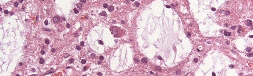
In Tumours, the normal control mechanisms of cell growth and proliferation are usually defective. One of the fundamental difference between tumours in Childeren and adults is that the nervous system in childeren are still developing and proliferation, whereas in adults, not all cells retain this abillty (like brain nerves). For this reason tumours of the nerves are found in young childeren, but not in adults , who have tumours in the supporting cells.
Some brain tumours destroy the brain tissue in which they grow. Some infiltrating tumours do this by depriving nutrients from the normal cells in the brain; some do this by exerting pressure by a growing mass. The brain functions associated with the involved regions are consequently lost. In order to understand the effect of tumours on the CNS, here is a short summary of some of the functions of the nervous system.
Nervous System
The nervous system detects features of the internal and external environment and in order to process these informations, nerve cells exist to tranfer these information/response rapidly from one part of the body to another. The nervous system is composed of nerves and ganglia's and also neuroglia's. The nerves enters and leaves the cranium through foramina’s. The nervous system (NS) has two major anatomical subdivisions:
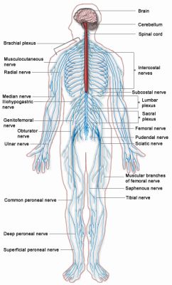
Image courtesy of Wiki Commons under creative commons license: https://commons.wikimedia.org/wiki/File:Nervous_system_diagram.png
1) Central nervous system (CNS); consists of the brain and the spinal cord, which are enclosed and protected by the cranium and vertebral column.
2) Peripheral nervous system (PNS) includes of all the nerves in the body, except the ones in the brain and spinal cord. It consist of afferent nerves, which conduct sensory information from effector cells to spinal cord and the brainstem. Efferent nerves transmit nerve impulses (response) from spinal cord and brain stem to target cells. Damage to a single nerve here results in weakness of the target muscle and loss of sensation of that region.
The brain
The average brain weight about 1.6kg and its size is proportinal to body size. The brain is divided into four major portions:
- Cerebrum
- Cerebellum
- Diencephalon
- Cerebrum
|
Image courtesy of Wiki Commons under creative commons license: https://commons.wikimedia.org/wiki/File:Gray728.svg |
Cerebrum
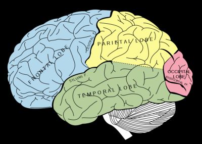
It’s the biggest of the four, constituting ~83% of the brain volume. It consist for two hemispheres (left and right) and these are separated from each other by the longitudinal fissure. Each hemisphere are composed of four major lobes: frontal, parietal, temporal, and the ocipital lobes and each has thick folds and shallow grooves (called gyri and sulci respectively).
- Frontal lobes: required for our personality, conscious thought and emotional control. Left lobe involved with language whereas right lobe involved in non-verbal abilities. Damage to the frontal lobe could cause a loss of ability to solve problems and to plan and initiate actions
- Parietal Lobes: concerned with cognition information processing, pain and touch sensation, spatial orientation, speech, visual perception. Damage to parietal lobe produce different symptoms depending on which hemisphere. For example, a lesion to left parietal lobe causes produces Gerstmann's Syndrome (neurological disorder characterised by different symptoms: such as writing disability (agraphia or dysgraphia), and an inability to distinguish right from left).
- Temporal lobes: concerned with hearing, smell and emotional behaviour. It also contains the hippocampus which is involed in memory processing. Damage to the temporal lobe causes disturbance of auditory sensation and perception, impaired long-term memory, Expressive aphasia (Broca's area 44&45), Receptive aphasia (Wernicke’s aphasia- disturbance of language comprehension), etc.
- Occipital lobe: principle visual centre of the brain. Damage causes visual hallucinations, blindness to one or both eyes.
Cerebellum
Also called the 'little brain',and consist of right and left cerebellar hemispheres. Cerebellum is mostly concerned with muscular co-ordination voluntary motor movement, balance and equilibrium and muscle tone. Damage leads to loss of coordination of motor movement (asynergia), inability to judge distance and when to stop (dysmetria), movement tremors (intention tremor), ect.
Diencephalon
Contains the thalamus, hypothalamus and the epithalums. They are involved in memory and emotional function and and homeostatic regulation.
Spinal Cord
Rounds from the base of the cranuim to the pelvic area. Spinal cord contain 31 pairs of spinal nerves; 8 servical (C1-C8), 12 thoracic (T1-T12), 5 lumbar (L1-L5), and 1 pair of Coccygeal nerves. every spinal nerve has two point of attachments to the spinal cord, Dorsal root, and ventral root.
Each spinal nerve (except C1) receives an sensory input from a specific area of t he skin called dermatomes, and one dermatome overlaps both neighbouring areas. Therefore damage to one of the spinal nerve will not produce a lost of sensation to that dermatome, as two neighbouring ones also innervate it.