Diagnosis
First point about cancer/tumour diagnosis is that there are no absolute methods for diagnosing and assessing the degree of malignancy of the tumour/cancer. Fortunately there several methods/ techniques those physicians/clinicians can use in order to identify the tumours. But first some symptoms of Tumours in CNS:
Symptoms of Tumour of the CNS
Tumour of the CNS are usually diagnosed when they produce and effect (i.e. black-outs, paralysis, loss of memory, even coma, etc). This effect could by caused by position and size of the tumour, which may press on or block a particular region of the central nervous system. For example, brain tumours (e.g. in choriod plexes) may cause a block of the cerebrospinal fluid (CSF), resulting in increase build up of fluid within the cranium, causing e.g. brain ischemia.
Microscopy
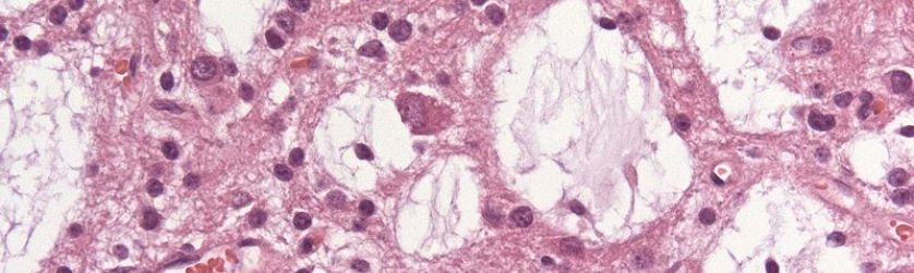
Pathologists routinely use is the microscopic examination for tumour diagnosis. They can look at a piece of tissue that has been biopsied, and they’ll have to make sound judgment (from experience) on whether a cell a sufficiently differentiated from the normal ones in order to classify it as a tumour cell. There two techniques used for tumour diagnosis:
- Tumour grading: this method measure using histological and cytological criteria degree of differentiation in tumours
- Tumour staging: this technique assesses the extent of tumour cell differentiation by using biological markers
Imaging
There are different imaging apparatuses that can be used to diagnose tumour of the CNS:
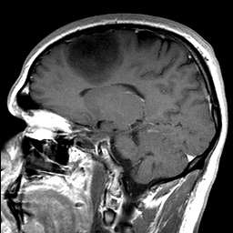
MRI (Magnetic Resonance imaging)- is machine that can safely produce imaging of high resolution of all tissues of the body. It has some pro’s and con’s, for example MRI emit no radiation, have a range of contrast, but they also very expensive and are slow (takes 10-50 minutes for an image) and has certain restrictions
|
image courtesy of Flickr under creative commons license::https://www.flickr.com/photos/46108355@N00/153101126/ |
CT scan- Is able to produce an image in less than 10 seconds. The image can differentiate between parenchyma and fluid such as CSF, and it can also show intera-cranial tissues. Pro’s and con’s of CT scan is that they cheap (compared to MRI), are widely available, and are not invasive. But they also produce radiation, show poor grey/white resolution and also produce artifact.